.jpg)

Assessment of Dental Aerosols Within a Dental Clinical Unit: An Avenue for the Transmission of Resistant Nosocomial Infection.
Sonia T Bredenkamp1*  and Charlene WJ Africa2
1Faculty of Dentistry, University of the Western Cape, Cape Town, South Africa .
2Department of Medical Biosciences, Faculty of Science, University of the Western Cape, Cape Town South Africa .
http://dx.doi.org/10.12944/EDJ.06.0102.10
The generation of aerosols in the dental clinic presents a risk for the transmission of nosocomial infections and should be monitored. Antimicrobial-resistant bacteria transmitted via routine dental procedures may complicate prophylaxis for infectious endocarditis. This study examined the role of dental aerosols in transmitting antimicrobial-resistant bacteria between dental healthcare personnel and patients in a dental teaching clinic. Using passive sampling, sterile blood agar plates were positioned at predetermined sites and exposed for 1 h after which they were incubated at 37 °C for 48 h. Their colony forming units (CFU) were quantified, and their species and antimicrobial susceptibility assessed using the VITEK®2 automated system. Compared with the control plates, plates exposed during scaling and polishing produced higher CFU counts than other restorative procedures. Gram-positive cocci constituted 104 (87%) of the total number of isolates (n = 119) with Micrococcus luteus (31.1%) and Staphylococcus hominis (8.4%) predominating Of ten strains of Staphylococcus hominis ssp hominis, all showed susceptibility to gentamycin, ciprofloxicin, clindamycin, linezolid, teicoplanin, vancomycin, tigecycline, rifampicin and trimethoprim/sulfamethoxazole; five showed resistance to fosfomycin; three showed resistance to erythromycin; and one showed resistance to oxacillin, daptomycin, tetracycline and fusidic acid. A low risk for the nosocomial transmission of resistant bacteria via aerosols was established.
Copy the following to cite this article:
Bredenkamp S. T, Africa C. W. J. Assessment of Dental Aerosols Within a Dental Clinical Unit: An Avenue for the Transmission of Resistant Nosocomial Infection. Enviro Dental Journal 2024; 6(1,2).
DOI:http://dx.doi.org/10.12944/EDJ.06.0102.10Copy the following to cite this URL:
Bredenkamp S. T, Africa C. W. J. Assessment of Dental Aerosols Within a Dental Clinical Unit: An Avenue for the Transmission of Resistant Nosocomial Infection. Enviro Dental Journal 2024; 6(1,2).Avialable here:https://bit.ly/3WhmAqv
Download article (pdf) Citation Manager

Introduction
The spread of nosocomial infections in healthcare settings poses a serious threat to public health, particularly in the dental clinic, where a high rate of nosocomial infections (infections notpresent when the patient was admitted) is known to occur.1 This may be attributed to several factors including the environment, patient immunosuppression, prolonged antibiotic use and inadequate practices among healthcare workers.2
There is a high risk of disease transmission in dentistry due to procedures that generate bioaerosols and splatter, 3-5 particularly in university settings where patients are attended to simultaneously. The use of high-speed cutting tools may result in an overload of contaminating aerosols.6
Pathogen transmission between patients and staff can occur through saliva, blood (especially accidental punctures with contaminated needles), inadequate surface and instrument sterilization (including dental unit waterlines)7-9 and the environmental dispersing of oral and respiratory tract microorganisms in dental aerosols and splatter.10 Aerosols generated by water and air instruments (aerosol particles of?11 and initiate a lower respiratory tract infection. Small particles do not settle quickly and their extended suspension in the air allows for their dispersal over a wider area, thereby increasing their potential to infect.12,13
Aerosol splatter are spread throughout the dental clinic and towards the dentist’s face and patient’s chest, respectively.9 Thoracic and inhalable aerosols (10–15 µm and 100-200 µm, respectively) penetrate the trachea and nasal areas, while droplets (>200 µm) are not airborne for extended periods and evaporate before reaching the ground or settling on the skin and mucous membranes of oral healthcare workers, resulting in contact infections.14 During the SARS- CoV-2 (also known as COVID-19) pandemic in 2019, infection control precautions were enhanced in all medical facilities to prevent the spread of the virus, directing the focus of healthcare workers to the role of aerosols in infection transmission.15,16
Despite creating different mitigation strategies and devices to address safety in aerosols and splatter generation17-19 most studies focused on viral aerosole contamination 20 and few studies reported on the monitoring and removing of microbiologically contaminated air from private and public dental rooms.21 The increased use of disinfectants during the COVID era contributed to an increase in the transmission of antimicrobial resistance (AMR) 22 with aerosols playing a major role in transmitting AMR genes23, 24 The United Nations Frontiers report 25 listed antimicrobial resistance as a significant growing public health concern and an understanding of the airborne transmission of AMR is essential because of its importance within the “One Health” concept. Even though their guidelines for dental practices were revised during the pandemic,26 neither the Centres for Disease Control (CDC) (2003) nor the World Health Organisation (WHO) guidelines27 mention the risk of AMR transmission via dental aerosols. The transmission of microbes during dental procedures should not be underestimated. The role of oral pathogens in the development of endocarditis during certain dental procedures has gained new interest, with Staphylococcus, Streptococcus and Enterococcus species contributing to approximately 90% of dentally-acquired infectious endocarditis cases.28 Increasing reports of their emerging AMR question the efficacy of currently used prophylactic antibiotics to prevent possible endocarditis.29 This study aimed to assess microbial dental aerosol transmission during basic restorative procedures to determine whether AMR bacteria can be nosocomially transmitted within a university dental clinical setting. To our knowledge, it is the first such study conducted in the Western region of XXXX with the following objectives:
To quantitatively and qualitatively assess dental aerosols before and during basic dental procedures using passive air sampling (using exposed blood agar plates) and
To identify the bacterial isolates and their antimicrobial profiles
Materials and Methods
Ethical approval
Ethical clearance was obtained from …XXXX…
Study design and area
This randomised clinico-microbial study consisted of microbial monitoring of the clinical environment in the restorative undergraduate dental clinic of XXXX. The clinical area is divided into eight dental cubicles. Each cubicle measures 696 m2, 2 960 m long, 2 440 m wide and 15 330 m high.
Microbial identification and characterization were performed in the laboratories of the Faculty of Natural Sciences in collaboration with …XXXX …
Clinical protocol
Aerosols were collected within the dental clinic using passive sampling. The standard booking protocol for the undergraduate dental clinic in the faculty of Dentistry required that service-rendering dentists screened the patients and recorded their demographics, medical history and complaint that brought them to the clinic and then referred them for treatment according to their treatment needs. Patients needing restorations and a scale and polish (n = 40) were referred to the primary investigator.
Microbial monitoring of aerosols was conducted on two to three patients/day during basic restorative work over a three-week period.
Anterior and posterior restorations included cavity preparation using a high-speed handpiece (Alegra TE98, Jean Weber and Hugo Hampel, Germany), caries removal using a low-speed handpiece (MKdent/Ecoline/ LE11, MK-dent), and an ultrasonic scaler (SONICflex 2000N /M161707, Kavodental) was used for scaling and polishing.
Aerosol Collection
The index of microbial air contamination (IMA) was determined using passive sampling on Columbia agar (Thermo Scientific, CM0331) supplemented with 5% sterile defibrinated horse blood (MRC horse farm, Cape Town).30 Blood agar (BA) plates were positioned on the counter and operational tray (Fig. 1) 1 m from the floor and 1 m away from the walls30 at a distance of 40 cm (between the tray and operatory site and between the counter and tray). BA plates were exposed to air for 1 h during the procedures then collected and incubated at 37 ºC for 48 h.21 A BA plate placed on the boundary wall in the clinic 1 h before the commencement of passive sampling on each sampling day served as a control.
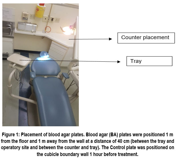 | Figure 1: Placement of blood agar plates. Blood agar (BA) plates were positioned 1 m from the floor and 1 m away from the wall at a distance of 40 cm (between the tray and operatory site and between the counter and tray).
|
Assessment of colony forming unis (CFU) on BA plates
Bacterial colony counts were recorded (Gallenkamp20//CX-300, Gallenkamp Co.Ltd., UK) and their morphology described.
Identification and characterisation of bacterial isolates
Pure cultures were obtained by subculture onto Trypticase soy agar (TSA, RMR0004, Merck Life Sciences GmbH, The Biovac Institute) and incubated for 24–48 hours at 37 ?.
BA plates were inspected for purity and confirmed by microscopic examination of Gram-stained smears at 100X magnification. Species were identified and their antimicrobial susceptibility determined using the VITEK® 2 system (bioMerieux Inc. France).
Using a sterile swab/ applicator stick, a colony was transferred from the pure growth on the TSA into the saline-containing polystyrene tube to adjust the turbidity to the required McFarland range (Table 1) using the DensiCHECKTM instrument (SKU93059, BioMerieux).
Table 1: The VITEK ® card turbidity range of bacterial suspensions
Product | McFarland Turbidity Range |
Gram-negative fermenting and non-fermenting bacilli | 0.50-0.63 |
Gram-positive cocci and non-spore-forming Bacilli | 0.50-0.63 |
Yeasts and yeast-like organisms | 1.80-2.20 |
Gram-positive spore-forming bacilli | 1.80-2.20 |
Reproduction from the the DensiCHECKTM package insert.
The appropriate VITEK® AST cards (AST-N255 and AST-P603 (bioMerieux, US) were selected for susceptibility testing according to the Gram stain morphology.
Statistical Analysis
Using ANOVA power analysis, it was estimated that 40 patients would be needed to obtain a statistically viable result for hypothesis testing.
The data were analyzed using IBM SPSS version 26. P < 0.05 indicated statistical significance.
Because their distribution was significantly non-normal, CFU distributions were compared to the location, dental treatment and student activity. Two independent samples were compared using the non-parametric Mann-Whitney test31 and more than two independent samples were compared using the Kruskal-Wallis test.32 Box and whisker plots demonstrated the group distributions.
Results
Calculating the Index of Microbial Air Contamination
Each location presented with a median of 5 (Table 2) demonstrating no significant difference between the distribution of colony counts in the control and each of the counter and tray locations (p = 0.881)
Table 2: A comparison of colony counts on blood agar before and after treatment exposure
Plate Location | Colony Forming Units/ Blood Agar Plate | |||||
Before Treat- ment Median | After treatment | |||||
Number of plates | Treatment | Median | Mean (SD) | P-value* | ||
Control (n = 19) | 5 | 19 | No treatment | 5 | 5.6 (3.7) | IA** |
Counter (n = 40) | 5 | 4 | Anterior restorations | 4.0 | 4.3 (3.3) | 0.360 |
9 | Posterior restorations | 4.0 | 4.4 (4.0) | |||
27 | Scale +polish | 5.0 | 7.5 (7.7) | |||
13 | All restorations | 4.0 | 4.4 (3.6) | |||
Tray (n = 40) | 5 | 4 | Anterior restorations | 4.5 | 4.3 (1.0) | 0.475 |
9 | Posterior restorations | 5.0 | 5.0 (3.5) | |||
27 | Scale + polish | 6.0 | 8.2 (7.7) | |||
13 | All restorations | 5.0 | 4.8 (2.9) | |||
*Statistical significance was defined as p < 0.05 (Kruskall-Wallis test) for comparison between dental treatments
**IA = no comparison groups were tested
Abbreviations: No=no treatment; AR=anterior restorations; PR=posterior restorations; ALL= all restorations; S+P= scale and polish
In summarising the descriptive statistics for the CFUs on the test plates (their location and number) and the dental treatment administered at the time of plate exposure (Table 2), we found that posterior restorations produced a higher CFU/plate count than anterior restorations on the tray plates.
No significant differences were observed between the counter (p = 0.360) and tray (p = 0.475) placements during different dental treatments as demonstrated in the Kruskal-Wallis test (Table 2).
Although the colony distributions of the control and counter placements were similar (Fig. 2) the tray placement CFUs were higher. However, this was not statistically significant (p = 0.881) as shown by the Kruskal-Wallis test.
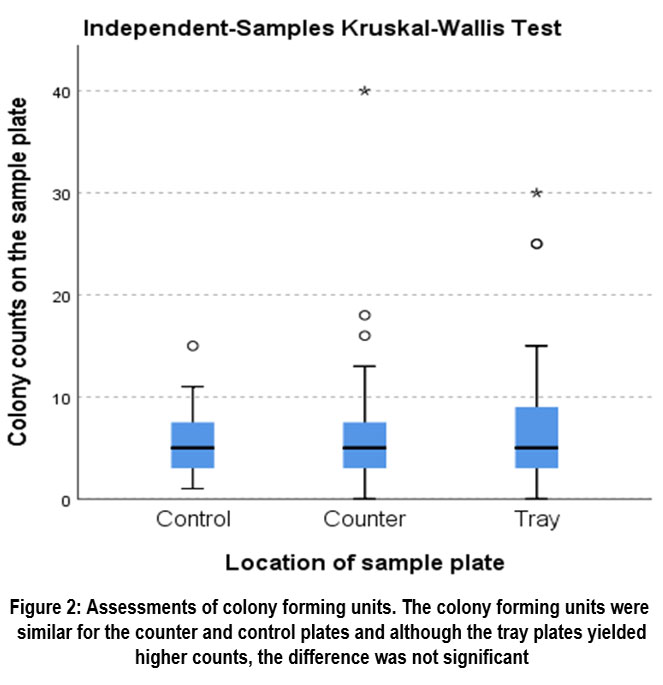 | Figure 2: Assessments of colony forming units. The colony forming units were similar for the counter and control plates and although the tray plates yielded higher counts, the difference was not significant
|
Isolation and identification of colonies
The total number of isolates comprised 119, with gram-positive species predominating and few gram-negative species isolated (Table 3).
The highest colony counts were observed on plates placed on the tray and following scaling and polishing exposure (Table 3).
Table 3: Frequency and percentage distribution of bacterial morphotypes according to dental procedures and plate locations.
Gram stain morphology | Dental treatment | Location of plate | Total n (%) | |||
Control n (%) | Restorations n (%) | Scale and Polish n (%) | Counter n (%) | Tray n (%) | ||
G-bacilli | 0 (0.0) | 0 (0.0) | 1 (1.5) | 0 (0.0) | 1 (1.8) | 1 (0.8) |
G-cocci | 1 (4.2) | 1 (3.4) | 1 (1.5) | 1 (2.5) | 1 (1.8) | 3 (2.5) |
G+bacilli | 4 (16.7) | 0 (0.0) | 7 (10.6) | 0 (0.0) | 7 (12.7) | 11 (9.2) |
G+cocci | 19 (79.2) | 28 (96.5) | 57 (86.3) | 39 (97.5) | 46 (83.6) | 104 (87.3) |
Total | 24 (100) | 29(100) | 66 (100) | 40 (100) | 55 (100) | 119 (100) |
G+ = gram-positive, G- = gram-negative
VITEK® identification of isolates
VITEK successfully identified 90% of the isolates, 4.2 % could not be identified and 5% had no or low reactive biopatterns (Supplementary Table A).
The predominant gram-positive cocci isolated were Micrococcus luteus (31%), Staphylococcus hominis ssp hominis (8.4%) and Kocuria rosea (7.6%). Among the other gram-positive cocci identified were the genera Aerococcus, Alloiococcus, Dermacoccus, Gamella, Globicatella, Granulicatella, Lactococcus and Leuconostoc (Supplementary Table A). Erysipelothrix and Rothia represented the gram-positive bacilli and none of the gram-negative cocci or bacilli were successfully identified.
Micrococcus luteus predominated regardless of the location (30% for the counter and 30.9% for the tray), student activity (24.1% versus 36.9% for student activity/ no student activity), and dental treatment procedure (37.9% for scale and polish and 27.3% for restorations). A similar trend was observed for each of the isolates identified (Supplementary Table A).
VITEK® Susceptibility testing
The bioMérieux instructional sheet (Ref 421040) for the VITEK® AST card guided the susceptibility testing of enterobacteriaceae, non-fermenters, staphylococci, enterococci, streptococci (including Streptococcus pnuemoniae, Streptococcus viridans, beta-haemolytic streptococci) and yeasts.
Antibiotic profiles are presented as S (susceptibility), R (resistance) or I (intermediate). Only S or R for a particular species indicates that all isolates of that species were susceptible or resistant to that particular antibiotic (Supplementary Table B).
Alloiococcus otitis species were susceptible to all the antibiotics tested and resistant to fosfomycin and fusidic acid (Supplementary Table B). Leuconostoc mesenteroides ssp cremoris was susceptible to all the antibiotics except erythromycin.
All four strains of Staphylococccus cohnii ssp cohnii were susceptible to gentamycin, ciprofloxacin, tigecycline and, trimethoprim/sulfamethoxazole; two strains were resistant to oxacillin and one strain was resistant to erythromycin, clindamycin, linezolid, daptomycin, teicoplanin, vancomycin, tetracycline, fosfomycin, fusidic acid and rifampicin. Three strains were resistant to fusidic acid, with only one intermediate result shown with rifampicin. Staphylococcus capitis was susceptible to all the antibiotics except fosfomycin. Staphylococcus cohnii ssp urealyticus was susceptible to most antibiotics and resistant to oxacillin and fusidic acid; intermediate susceptibility was shown to clindamycin and tetracycline. Staphylococcus epidermidis was susceptible to most antibiotics except oxacillin, erythromycin, clindamycin, tetracycline and trimethoprim/sulfamethoxazole. All Staphylococcus hominis ssp hominis strains were susceptible to gentamycin, ciprofloxicin, clindamycin, linezolid, teicoplanin, vancomycin, tigecycline, rifampicin and trimethoprim/sulfamethoxazole; five were resistant to fosfomycin; three were resistant to erythromycin; and one was resistant to oxacillin, daptomycin, tetracycline and fusidic acid. Staphylococcus saprophyticus was resistant to all the antibiotics except erythromycin, fusidic acid and fosfomycin. Streptococcus mitis/oralis and Streptococcus salivarius ssp salivarius were susceptible to vancomycin, ampicillin, penicillin and clindamycin.
Discussion
Passive air sampling reflects the extent of contamination on critical surfaces at the end of a treatment session and highlights high-risk areas for contamination between patients.33 The present study used passive air sampling to assess dental aerosols within a university dental clinical when routine dental procedures (fillings and scaling) were performed. Further, it determined the antimicrobial profiles of the bacterial isolates, to establish whether a risk for nosocomial transmission of antimicrobial-resistant bacteria could occur within a dental clinical setting.
No significant difference was found between the baseline colony counts for the control plates and the tray/ counter placements, although the colony counts for the tray placements were higher than the counter placements, unlike the study by Zemouri et al 34 that showed a significantly lower IMA in the dental clinic before treatment. The higher colony counts for the tray placements concur with the studies of Madhuri and Girish Babu 35 and Zemouri et al 34 where the highest CFU’s were concentrated at the chest and instrument area. Yang et al 1 also found the highest level of contamination in a triangle formed between the chest area of the dentist, dental assistant and patient. The patient’s chest area was the most heavily contaminated area.15
As previously reported,1,36-37 scaling and polishing generated higher colony counts than anterior and posterior restorations followed by cavity preparation using high speed handpieces. By contrast, Matys and Grzech-Lesniak,8 indicated that less aerosol was generated through ultrasonic scaling compared to the use of rotary instruments. This was also a key finding in a recent systematic review.38
A comparison of the higher mean CFU/plate for posterior and anterior restorations may be due to the fact that more time was needed on the complex depth required for the preparation of posterior restorations, with more aerosol from the water spray influencing the risk of increasing microbial transmission.8 Earlier clinical studies of dental aerosols included dental procedures as part of their study design, but their methodologies differed according to the objectives and we were unable to support or negate their findings.39-43
The distance of aerosol spread and their level of contamination in the dental surgery is a matter of concern, especially with the number of patients with oro-nasal methicillin-resistant Staphylococcus aureus (MRSA) steadily increasing. This, in addition to the emergence of new pathogens, increasing AMR, the introduction of faster-rotating instruments, modern devices and equipment (often into outdated facilities and settings), present their own challenges.44
The wide distribution of aerosols that remain suspended for a considerable time in dental clinics before settling,45 increase the risk of exposure, thereby increasing the risk of contact infection as well as airborne infection.12,13-,45 Cross-contamination is inevitable in crowded spaces even though transmission may not necessarily result in infection.
A distance of more than six feet (182.88 cm) away from a patient infected with COVID was considered a risk of disease transmission via aerosols.12,46 Considering that aerosols may travel a longer distance before settling and continue to remain airborne for 30 minutes after the dental procedure,47 the placement of plates at 40 cm could thus be perceived as a limitation of the present study. However, as the particle size increases (bacteria are larger than viruses), the distance travelled is shorter,48 and thus we consider the placement distance adequate for meeting the objectives of this study (to detect aerosol bacterial contamination). The systematic assessment and monitoring of dental aerosol to prevent transmission of infection is important,49 and this is one of a paucity of studies conducted in Africa 50 in the past decade.
The low yield of our findings is reassuring and indicates that dental aerosols do not pose a significant health hazard in transmitting AMR nosocomial infections in the dental clinic. Most studies of aerosol-generating procedures (AGP) use settle plates to detect droplets that have fallen, 10,15,51 even though settle plates may not capture aerosols that persist for longer periods, or the transmissibility of viruses.52
VITEK® technology was used for identifying the species and antimicrobial profiles of 119 species in this study. VITEK® technology is widely used to identify clinical and environmental microorganisms, and to determine microbial susceptibility to antibiotics with accurate and conclusive reliability,37,41 with the only limitation being attributed to the capacity of the system’s database.53 As in other studies,34,39,54 a variation in the bacterial composition of aerosols in the dental clinic was observed comprising a mixture of species from the environment, skin and oral cavity. Although few in number, less-reported antimicrobial resistant species were identified, making this an important contribution to the current literature.
As indicated above, the predominant isolates were gram-positive bacteria and included the genera Micrococcus, Staphylococcus, and Streptococcus. Gram-positive bacilli previously reported included the genera Actinomyces, Bacillus and Corynebacterium.12,34,37,38,39,40,42,54
The predominant species in this study were Micrococcus luteus and S. homini, both of which have been reported in rare cases of infectious endocarditis and bacteraemia associated with dental procedures.55,56 Micrococci are found in abundance on the lingual surfaces intra-orally especially on the surface of the tongue,39 while S. hominis has been reported as a potentially pathogenic bacterium found in the human oropharynx and upper respiratory tract.57 Staphylococcus aureus was not isolated from any of the plates in this study, and its absence coupled with the prominence of coagulase-negative staphylococci (CoNS) such as S. hominis, has been reported in other studies of airborne bacteria in dental clinics.58
Other gram-positive cocci isolated in this study that are rarely considered pathogenic and are under-reported but have been implicated in nosocomially-acquired endocarditis include Granulicatella, 59, Lactococcus lactis, 60 and Gamella species 61. Among the gram-positive cocci, AMR was most prevalent in the CoNS bacteria with several species showing resistance to oxacillin, erythromycin, tetracycline, clindamycin, vancomycin, fucidin and fosfomycin as previously reported.62
Pseudomonas aeruginosa, S. aureus, Klebsiella spp, Enterococcus spp, Escherichia coli and CoNS are among the most common AMR nosocomial medical and dental pathogens63,64 with several species included in the consortium of ESKAPE pathogens (Enterococcus faecalis, Staphylococcus aureus, Klebsiella pneumoniae, Acinetobacter baumannii, Pseudomonas aeruginosa, and Enterobacter) that have gained interest in Dentistry, since they are frequently associated with post-operative or opportunistic infections.65 Their antimicrobial resistance complicates effective antibiotic treatment and adversely affects the treatment of severely infected patients.66,67 K. pneumoniae, P. aeruginosa, A. baumannii, and S. aureus have been reported in infections in the oral and maxillofacial regions,67,68 creating a need for further exploratory studies of the potential risk for nosocomial infection within the dental setting 15,49 compared to the medical setting.
WHO has declared AMR as one of the top 10 global public health threats .69 Studies in the UK70 and America 71 found that 80% of antibiotics prescribed for treatment and prophylaxis respectively, were inappropriate or totally unnecessary. Therefore, dental professionals should strive to reduce and prevent the indiscriminate use of antibiotics to reduce the risks of AMR.
Among the limitations of the study are:
A failure to recover Klebsiella or other enteropathic bacteria in the present study, may suggest that they are largely transmitted via direct contact and are not airborne. An examination of the gear worn by the dentist may have confirmed this and may be perceived as a limitation of the study. However, that was outside the scope of this study, and we believe that we have achieved the aim of the study which was to compare the airborne bacteria before and after treatment.
Failure to incubate the plates under anaerobic or capnophilic conditions may also be perceived as a limitation, since previous studies have listed Actinomyces and Capnocytophaga as anaerobic organisms isolated from dental aerosols 34 and the HACEK group of bacteria (Haemophilus spp., Aggregatibacter actinomycetemcomitans, Cardiobacterium hominis, Eikenella corrodens, and Kingella kingae) have been implicated in the nosocomial transmission of infectious endocarditis.72 Although these genera grow better in anaerobic or capnophilic environments, some are aerotolerant and can grow on BA under facultatively anaerobic conditions as well, so their isolation would not have been missed in the present study. We considered the use of BA plates appropriate for this purpose since BA supports the growth of aerobes and facultative anaerobes, including gram-negative bacilli that are often selected for on MacConkey agar plates. However, the oral obligate anaerobes are extremely fastidious and would not have survived exposure to air for an hour, thus we did not consider anaerobic incubation of the exposed plates. However, we concede that settle plates may not provide an accurate presentation of the microbial composition of the aerosols in that there is a bias towards facultative anaerobes and aerobes and that DNA isolation and sequencing may prove more efficient.
Conclusion
The results of this study demonstrated that the index of microbial contamination was acceptable at baseline and during dental treatment and showed no risk for the transmission of AMR pathogens.
The empiric antibiotics most frequently used for the oral and maxillofacial area are amoxicillin-clavulanic acid, metronidazole, and erythromycin; however, the number of antibiotic-resistant bacteria is rapidly increasing, and therefore the dental team in close collaboration with pharmacists, microbiologists, and other health care professionals, should work together to assure an antibiotic stewardship programme and stay abreast of AMR.
Species identification and antimicrobial profiles of bacterial isolates are important because antibiotics are prescribed by dentists for prophylaxis to prevent infectious endocarditis, to improve the outcome success of surgical interventions and to treat current oral infections. VITEK® and other automated technologies facilitate the process of regular environmental monitoring. Air quality analysis is an important component of environmental microbiological monitoring to determine the risk of transmitting AMR airborne infectious pathogens, especially in enclosed environments such as healthcare facilities. The scope for future studies should include consistent and systematic monitoring of nosocomial infections within the dental setting especially within the open-plan clinical training platform in dental schools. Acknowledging the inter-disciplinary nature of the “One Health” framework will provide a better picture of the spread of AMR through environmental monitoring, thereby presenting a new dimension beyond just the inappropriate use of antimicrobials. This will inform more effective mitigation measures to reduce microbial air contamination and control the aerosole transmission of nosocomially-acquired AMR.
Acknowledgement
The authors acknowledge the collaborative contributions of the following institutions: the Faculty of Applied Sciences, Cape Peninsula University of Technology; AMPATH laboratories, Cape Town; and the bioMérieux Applications Specialist. We gratefully acknowledge Pedro Abrantes, Vanessa Jooste, and Rose Gwanpu Epse Gwanfogbe for assistance in the laboratory, and Tonya Esterhuizen at the University of Stellenbosch for statistical analysis
Funding Sources
This work was supported by the National Research Foundation [grant number: 129321].
Conflict of Interest
The author(s) do not have any conflict of interest.
Data Availability Statement
All data are provided in the manuscript and supplementary tables. Any additional data can be provided upon reasonable request from the corresponding author.
Ethics Statement
This research did not involve human participants, animal subjects, or any material that requires ethical approval.
Informed Consent Statement
Because routine dental treatment was carried out according to normal clinical protocol, patient consent was not required, although patients were informed that sample plates would be placed within the cubicle and that the data collected would be used for research purposes and kept confidential.
Clinical Trial registration
Not applicable. This research does not involve any clinical trials.
Permission to Reproduce Material from Other Sources
Not Applicable
Author Contributions
Sonia Bredenkamp: Conceptualisation, Data curation, Formal analysis, Investigation, Methodology, Validation, Visualization, Writing - original draft.
Charlene Africa: Methodology, Project administration, Resources, Supervision, Validation, Writing, review & editing.
References
- Yang M., Chaghtai A., Meléndez M., Hasson H., Whitaker E.J., Badi M.A., Mitchell J.C., Newsome A.D. Mitigating saliva aerosol contamination in a dental school clinic. BMC Oral Health. 2021; 21:1-10. doi:10.1186/s12903-021-01417.
CrossRef - Khan H.A., Baig F.K., Mehboob R. Nosocomial infections: Epidemiology, prevention, control and surveillance. Asian Pac J Trop Biomed. 2017; 7:478-482. doi: 10.1016/j.apjtb.2017.01.019.
CrossRef - Proffitt E. What will be the new normal for the dental industry? Br Dent J. 2020; 228:678-680. doi:10.1038/s41415-020-1583-x.
CrossRef - Abusalim GS. Prevalence and investigations of bacterial contamination in dental healthcare associated environment. J King Saud Univ Sci. 2022;34(6):102153. doi:10.1016/j.jksus.2022.102153.
CrossRef - Jain M, Mathur A, Mathur A, Mukhi PU, Ahire M, Pingal C. Qualitative and quantitative analysis of bacterial aerosols in dental clinical settings: Risk exposure towards dentist, auxiliary staff, and patients. J Family Med Prim Care. 2020;9(2):1003. doi:10.4103/jfmpc.jfmpc_863_19
CrossRef - Takanabe Y., Maruoka Y., Kondo J., Yagi S., Chikazu D., Okamoto R., Sakagami R., Nakamura T. Dispersion of aerosols generated during dental therapy. Int J Environ Res Public Health. 2021; 18:11279. doi:10.3390/ijerph182111279.
CrossRef - Laheij A.M.G.A., Kistler J.O., Belibasakis G.N., Välimaa H., de Soet J.J. Healthcare-associated viral and bacterial infections in dentistry. J Oral Microbiol. 2012; 4:17659. doi:10.3402/jom. v4i0.17659.
CrossRef - Matys J., Grzech-Le?niak K. Dental aerosol as a hazard risk for dental workers. Materials (Basel). 2020; 13:5109. doi:10.3390/ma13225109.
CrossRef - Yu Y., Wu X., Sun Y. Precise control of digital dental unit to reduce aerosol and splatter production: new challenges for future epidemics. BMC Oral Health. 2024; 24:1-8. doi:10.1186/s12903-024-03980-w.
CrossRef - Ionescu A.C., Cagetti M.G., Ferracane J.L., Garcia-Godoy F., Brambilla E. Topographic aspects of airborne contamination caused by the use of dental handpieces in the operative environment. J Am Dent Assoc. 2020; 151:660-667. doi: 10.1016/j.adaj.2020.06.002.
CrossRef - Lahdentausta L., Sanmark E., Lauretsalo S., Korkee V., Nyman S., Atanasova N., Pitkänen T., Jämsä T., Meurman J.H. Aerosol concentrations and size distributions during clinical dental procedures. Heliyon. 2022;8:e11074. doi: 10.1016/j.heliyon.2022.e11074.
CrossRef - Kumar P.S., Subramanian K. Demystifying the mist: Sources of microbial bioload in dental aerosols. J Periodontol. 2020; 91:10.1002/JPER.20-0395. doi:10.1002/JPER.20-0395.
CrossRef - Dudding T., Sheikh S., Florence F., Haworth J., Haworth S., Main B., Jinks S., Holliday R. A clinical observational analysis of aerosol emissions from dental procedures. PLoS One. 2022;17: e0265076. doi: 10.1371/journal.pone.0265076.
CrossRef - Van der Weijden F. Aerosol in the oral health-care setting: a misty topic. Clin Oral Investig. 2023; 27:23-32. doi:10.1007/s00784-023-05034-x.
CrossRef - Innes N., Johnson I.G., Al-Yaseen W., Harris R., Jones R., KC S., Longman L., Downer K., Bhagat R., Reay S., Wade W.G., Robertson M. A systematic review of droplet and aerosol generation in dentistry. J Dent. 2021; 105:103556. doi: 10.1016/j.jdent.2020.103556.
CrossRef - Noordien N., Mulder-van Staden S., Mulder R. Correction: Noordien et al. In vivo study of aerosol, droplets and splatter reduction in dentistry. Viruses. 2023; 15:1669. doi:10.3390/v15081669.
CrossRef - Comisi J.C., Ravenel T.D., Kelly A., Teich S.T., Renne W. Aerosol and spatter mitigation in dentistry: Analysis of the effectiveness of 13 setups. J Esthet Restor Dent. 2021; 33:466-479. doi:10.1111/jerd.12717.
CrossRef - Allison J.R., Dowson C., Pickering K., ?ervinskyt? G., Durham J., Jakubovics N.S., Holliday R. Local exhaust ventilation to control dental aerosols and droplets. J Dent Res. 2021; 100:002203452110562. doi:10.1177/00220345211056287.
CrossRef - Nulty A., Lefkaditis C., Zachrisson P., Van Tonder Q., Yar R. A clinical study measuring dental aerosols with and without a high-volume extraction device. Br Dent J. 2020; 228:1-8. doi:10.1038/s41415-020-2274-3.
CrossRef - Locke L, Dada O, Shedd JS. Aerosol transmission of infectious disease and the efficacy of personal protective equipment (PPE): A systematic review. J Occup Environ Med. 2021;63(11):e783-e791. doi:10.1097/JOM.0000000000002366.
CrossRef - Rosa V., Agarwalla S.V., Tan B.L., Choo S.Y., Sim Y.F., Boey F.Y.C., Goh C.L. Pandemic preparedness and response: A foldable tent to safely remove contaminated dental aerosols—Clinical study and patient experience. Appl Sci. 2022; 12:7409. doi:10.3390/app12157409.
CrossRef - Li J, Cao J, Zhu YG, et al. Global survey of antibiotic resistance genes in air. Environ Sci Technol. 2018;52(19):10975-10984. doi:10.1021/acs.est.8b02204.
CrossRef - Jin L, Xie J, He T, Wu D, Li X. Airborne transmission as an integral environmental dimension of antimicrobial resistance through the “One Health” lens. Crit Rev Environ Sci Technol. 2021;52(23):4172-4193. doi:10.1080/10643389.2021.2006537.
CrossRef - Wang Q, Liu C, Sun S, et al. Enhance antibiotic resistance and human health risks in aerosols during the COVID-19 pandemic. Sci Total Environ. 2023;871:162035. doi:10.1016/j.scitotenv.2023.162035.
CrossRef - United Nations Environment Programme (UNEP). Frontiers 2017: Emerging Issues of Environmental Concern. Nairobi, Kenya: United Nations Environment Programme; 2017.
- World Health Organization. Considerations for the Provision of Essential Oral Health Services in the Context of COVID-19: Interim Guidance. August 2020. Accessed July 14, 2025. https://www.who.int/publications/i/item/who-2019-nCoV-oral-health-2020.1
- World Health Organization. Infection Prevention and Control of Epidemic and Pandemic-Prone Acute Respiratory Infections in Health Care: WHO Guidelines. Published 2014. Accessed July 14, 2025. https://www.who.int/csr/bioriskreduction/infection_control/publication/en/
- Cahill T.J., Prendergast B.D. Infective endocarditis. Lancet. 2016; 387:882-893. doi:10.1016/s0140-6736(15)00067-7.
CrossRef - Komiyama E.Y., Lepesqueur L.S.S., Yassuda C.G., Samaranayake L.P., Parahitiyawa N.B., Balducci I., Koga-Ito C.Y. Enterococcus species in the oral cavity: Prevalence, virulence factors and antimicrobial susceptibility. PLoS One. 2016;11: e0163001. doi: 10.1371/journal.pone.0163001.
CrossRef - Pasquarella C., Pitzurra O., Savino A. The index of microbial air contamination. J Hosp Infect. 2000; 46:241-256. doi:10.1053/jhin.2000.0820.
CrossRef - Mann H.B., Whitney D.R. On a test of whether one of two random variables is stochastically larger than the other. Ann Math Stat. 1947; 18:50-60. doi:10.1214/aoms/1177730491.
CrossRef - Kruskal W.H., Wallis W.A. Use of ranks in one-criterion variance analysis. J Am Stat Assoc. 1952; 47:583-621.
CrossRef - Maghlouth A.A., Yousef Y.A., Bagieh N.A. Qualitative and quantitative analysis of bacterial aerosols. J Contemp Dent Pract. 2004; 5:91-100. doi:10.5005/jcdp-5-4-91.
CrossRef - Zemouri C., Volgenant C.M.C., Buijs M.J., Crielaard W., Rosema N.A.M., Brandt B.W., van der Weijden F. Dental aerosols: microbial composition and spatial distribution. J Oral Microbiol. 2020; 12:1762040. doi:10.1080/20002297.2020.1762040.
CrossRef - Babu R.J.G., M.K.R. Evaluation of bacterial aerosol contamination during dental procedures. IP Int J Med Microbiol Trop Dis. 2019; 5:23–7. doi:10.18231/2581-4761.2019.0006.
CrossRef - Alharbi A., Alharbi S., Alqaidi S. Guidelines for dental care provision during the COVID-19 pandemic. Saudi Dent J. 2020; 32:181–6. doi: 10.1016/j.sdentj.2020.04.001.
CrossRef - Boccia G., Di Spirito F., D’Ambrosio F., De Caro F., Pecora D., Giorgio R., Battista G. Microbial air contamination in a dental setting environment and ultrasonic scaling in periodontally healthy subjects: An observational study. Int J Environ Res Public Health. 2023; 20:2710. doi:10.3390/ijerph20032710.
CrossRef - Nóbrega M.T.C., Bastos R.T.R.M., Mecenas P., de Toledo I.P., Richardson-Lozano R., Altabtbaei K., Araújo L. Aerosol generated by dental procedures: A scoping review. J Evid Based Med. 2021; 14:303–12. doi:10.1111/jebm.12461.
CrossRef - Mathur A., Jain M., Mathur A., Mukhi P., Ahire M., Pingal C. Qualitative and quantitative analysis of bacterial aerosols in dental clinical settings: Risk exposure towards dentist, auxiliary staff, and patients. J Fam Med Prim Care. 2020; 9:1003. doi: 10.4103/jfmpc.jfmpc_863_19.
CrossRef - Mirhoseini S.H., Koolivand A., Bayani M., Sarlak H., Moradzadeh R., Ghamari F., Daryabeigi R. Quantitative and qualitative assessment of microbial aerosols in different indoor environments of a dental school clinic. Aerobiologia. 2021; 37:217–24. doi:10.1007/s10453-020-09679-z.
CrossRef - Choudhary S., Bach T., Wallace M.A., Stoeckel D.C., Thornhill M.H., Lockhart P.B., Seymour C.W. Assessment of infectious diseases risks from dental aerosols in real-world settings. Open Forum Infect Dis. 2022;9. doi:10.1093/ofid/ofac617.
CrossRef - Jyoti S. Microbial aerosols generation during various dental procedures in a university hospital. Al Ameen J Med Sci. 2022;15.
- Rafiee A., Carvalho R., Lunardon D., Flores-Mir C., Major P., Quemerais B., Lagravère M. Particle size, mass concentration, and microbiota in dental aerosols. J Dent Res. 2022; 101:785–92. doi:10.1177/00220345221087880.
CrossRef - Rautemaa R., Nordberg A., Wuolijoki-Saaristo K., Meurman J.H. Bacterial aerosols in dental practice—a potential hospital infection problem? J Hosp Infect. 2006; 64:76–81. doi: 10.1016/j.jhin.2006.04.011.
CrossRef - Takanabe Y., Maruoka Y., Kondo J., Yagi S., Chikazu D., Okamoto R., Takanashi K. Dispersion of aerosols generated during dental therapy. Int J Environ Res Public Health. 2021; 18:11279. doi:10.3390/ijerph182111279.
CrossRef - Klompas M., Baker M.A., Rhee C. Airborne transmission of SARS-CoV-2. JAMA. 2020; 324:441. doi:10.1001/jama.2020.12458.
CrossRef - Bennett A.M., Fulford M.R., Walker J.T., Bradshaw D.J., Martin M.V., Marsh P.D. Microbial aerosols in general dental practice. Br Dent J. 2000; 189:664–7. doi: 10.1038/sj.bdj.4800859a.
CrossRef - Demirkol N., Karagözo?lu ?., Koçer ?. Efficiency of HEPA-filtered extra-oral suction unit on aerosols during prosthetic dental preparation: A pilot study. Clin Oral Investig. 2023; 27:1937–44. doi:10.1007/s00784-022-04850-x.
CrossRef - Horzov L.F., Kryvtsova M.V., Kostenko Y.Y., Kalynyak M.M. Microbial composition of dental clinics' air as a factor of spread of nosocomial infections. Ukr J Med Biol Sports. 2020; 5:214–7. doi:10.26693/jmbs05.06.214.
CrossRef - Bayingana C., Gahutu J.B., Sendegeya A., Habarugira F., Mukumpunga C., Lyumugabe F., et al. Prevalence of bacterial species involved in nosocomial infections in pediatrics unit at Butare University Teaching Hospital (Chub). Int J Curr Microbiol Appl Sci. 2017; 6:3227–32. doi:10.20546/ijcmas.2017.606.380.
CrossRef - Garland K.V. A safe approach to aerosols. Dimens Dent Hyg. 2022;20.
- Johnson I.G., Jones R.J., Gallagher J.E., Wade W.G., Al-Yaseen W., Robertson M., et al. Dental periodontal procedures: a systematic review of contamination (splatter, droplets and aerosol) in relation to COVID-19. BDJ Open. 2021; 7:1–7. doi:10.1038/s41405-021-00070-9.
CrossRef - Al Bulushi I.M., Al Kharousi Z.S., Rahman M.S. Vitek: a platform for a better understanding of microbes. Tech Meas Food Saf Qual. 2021;117–36. doi:10.1007/978-3-030-68636-9_6.
CrossRef - Anjum A., Hosein M., Butt S.A., Fakhuruddin, Shafiq Y. Qualitative analysis of bacterial aerosols generated during ultrasonic dental scaling. J Adv Med Med Res. 2020; 31:1–12. doi:10.9734/jammr/2019/v31i1130333.
CrossRef - Becker K., Heilmann C., Peters G. Coagulase-negative staphylococci. Clin Microbiol Rev. 2014; 27:870–926. doi:10.1128/cmr.00109-13.
CrossRef - Vasconcellos D., Weng B.H., Wu P., Thompson G.M., Sutjita M. Staphylococcus hominis infective endocarditis presenting with embolic splenic and renal infarcts and spinal discitis. Case Rep Infect Dis. 2022; 2022:1–5. doi:10.1155/2022/7183049.
CrossRef - Szczuka E., Krzymi?ska S., Bogucka N., Kaznowski A. Multifactorial mechanisms of the pathogenesis of methicillin-resistant Staphylococcus hominis isolated from bloodstream infections. Antonie Van Leeuwenhoek. 2018; 111:1259–65. doi:10.1007/s10482-017-1007-3.
CrossRef - Aquino de Muro M., Shuryak I., Uhlemann A., Tillman A., Seeram D., Zakaria J., et al. The abundance of the potential pathogen Staphylococcus hominis in the air microbiome in a dental clinic and its susceptibility to far-UVC light. Microbiol Open. 2023;12. doi:10.1002/mbo3.1348.
CrossRef - Cargill J.S., Scott K.S., Gascoyne-Binzi D., Sandoe J.A.T. Granulicatella infection: diagnosis and management. J Med Microbiol. 2012; 61:755–61. doi:10.1099/jmm.0.039693-0.
CrossRef - Freires I.A., Avilés-Reyes A., Kitten T., Simpson-Haidaris P.J., Swartz M., Knight P.A. Heterologous expression of Streptococcus mutans Cnm in Lactococcus lactis promotes intracellular invasion, adhesion to human cardiac tissues and virulence. Virulence. 2016; 8:18–29. doi:10.1080/21505594.2016.1195538.
CrossRef - Ural S., Yurtsever S.G., Ormen B., Turker N., Kaptan F., El S. Gemella morbillorum endocarditis. Case Rep Infect Dis. 2014; 2014:1–3. doi:10.1155/2014/456471.
CrossRef - Chabi R., Momtaz H. Virulence factors and antibiotic resistance properties of the Staphylococcus epidermidis strains isolated from hospital infections in Ahvaz, Iran. Trop Med Health. 2019;47. doi:10.1186/s41182-019-0180-7
CrossRef. - MacGowan A., Macnaughton E. Antibiotic resistance. Medicine (Baltimore). 2017; 45:622–8. doi: 10.1016/j.mpmed.2017.07.006.
CrossRef - Zawadzki P.J., Perkowski K., Padzik M., Mierzwinska-Nastalska E., Szaflik J.P., Conn D.B. Examination of oral microbiota diversity in adults and older adults as an approach to prevent spread of risk factors for human infections. Biomed Res Int. 2017; 2017:1–7. doi:10.1155/2017/8106491.
CrossRef - Lee H.J., Moon S.Y., Oh J.S., Choi H.I., Park S.Y., Kim T.E. ESKAPE pathogens in oral and maxillofacial infections. J Oral Med Pain. 2022; 47:52–61. doi:10.14476/jomp.2022.47.1.52.
CrossRef - Ma Y.X., Wang C.Y., Li Y.Y., Li J., Wan Q.Q., Chen J.H. Considerations and caveats in combating ESKAPE pathogens against nosocomial infections. Adv Sci (Weinheim). 2020; 7:1901872. doi:10.1002/advs.201901872.
CrossRef - Kang S.H., Kim M.K. Antibiotic sensitivity and resistance of bacteria from odontogenic maxillofacial abscesses. J Korean Assoc Oral Maxillofac Surg. 2019; 45:324. doi:10.5125/jkaoms.2019.45.6.324.
CrossRef - Park S.Y., You J., Moon S.Y., Oh J.S., Choi H.G., Jung G.W. A literature review of infection with ESKAPE pathogens in oral and maxillofacial region. J Oral Med Pain. 2021; 46:75–83. doi:10.14476/jomp.2021.46.3.75.
CrossRef - World Health Organization. Antimicrobial Resistance. Updated 2024. Accessed July 14, 2025. https://www.who.int/news-room/fact-sheets/detail/antimicrobial-resistance
- Cope AL, Francis NA, Wood F, Chestnutt IG. Antibiotic prescribing in UK general dental practice: a cross-sectional study. Community Dent Oral Epidemiol. 2015;44(2):145-153. doi:10.1111/cdoe.12199
CrossRef - Suda KJ, Calip GS, Zhou J, et al. Assessment of the appropriateness of antibiotic prescriptions for infection prophylaxis before dental procedures, 2011 to 2015. JAMA Netw Open. 2019;2(5):e193909. doi:10.1001/jamanetworkopen.2019.3909
CrossRef - Dominiak M., Ró?y?o-Kalinowska I., Gedrange T., Konopka T., Hadzik J., Bednarz W. COVID-19 and professional dental practice. The Polish Dental Association Working Group recommendations for procedures in dental office during an increased epidemiological risk. J Stomatol. 2020; 73:1–10. doi:10.5114/jos.2020.94168.
CrossRef