.jpg)
Assessment of Mandibular Root Morphology Using Cone Beam Computed Tomography in the Croatian Population
Lucija Kustra1  , Ivana Medvedec Mikic2 and Kristina Gorseta3*
1Department of Pediatric and Preventive Dentistry, School of Dental Medicine, University of Zagreb, Zagreb, Croatia .
2Department of Endodontics and Restorative Dental Medicine, School of Medicine, University of Split, Split, Croatia .
3Clinical Hospital Center Zagreb, University Dental Clinic, Zagreb, Croatia .
http://dx.doi.org/10.12944/EDJ.08.0107
The objective of this research was to study the variation in the number of roots of mandibular teeth and to compare them with data from available literature. The retrospective study was conducted at the Department of Maxillo-facial Surgery at the University Hospital Centre Split by analysing Cone-Beam Computed Tomography (CBCT) scans of patients' lower jaws. Out of 145 CBCT scans of the lower jaws, 114 were valid and entered in the Excel table for statistical analysis. Three hypotheses have been put forward: (1) lower incisors, canines and premolars have one root; (2) lower molars have two roots; (3) supernumerary root is the most common variation in lower molars. Data collected from 114 participants, i.e. CBCT scans, were analysed in the STATISTICA 11.0 programme package and every side of a group of teeth was analysed separately. Lower incisors, canines and premolars are, in the largest percentage, single-rooted teeth (two-rooted teeth vary from 0 to 4 per cent), whereas molars are, the most, two-rooted teeth (one-rooted teeth vary from 2 to 5 per cent, while three-rooted teeth stand at about 3 to 6 per cent). The most frequent anatomical variation observed was the presence of a supernumerary root in the mandibular third molar. This research confirmed the hypotheses and obtained data on the number of dental roots in all teeth of the lower jaw, in accordance with data from the literature.
Copy the following to cite this article:
Kustra L, Mikic I. M, Gorseta K. Assessment of Mandibular Root Morphology Using Cone Beam Computed Tomography in the Croatian Population. Enviro Dental Journal 2026;8(1).
DOI:http://dx.doi.org/10.12944/EDJ.08.0107Copy the following to cite this URL:
Kustra L, Mikic I. M, Gorseta K. Assessment of Mandibular Root Morphology Using Cone Beam Computed Tomography in the Croatian Population. Enviro Dental Journal 2026;8(1). Avialable here: https://bit.ly/4cm8Kw8
Download article (pdf) Citation Manager

Introduction
Endodontics is a branch of dental medicine focusing on the morphology, physiology, pathology, diagnosis, and treatment of the dental pulp and periapical tissues. A thorough understanding of tooth anatomy, especially the endodontic area, is essential for high-quality dental treatment. Endodontic procedures involve creating access openings, instrumentation, and filling of root canals, aimed at preserving the tooth for as long as possible. Failure to locate root canal entrances, improper instrumentation, or inadequate filling often leads to treatment failure.1
Besides clinical examination, radiographic analysis plays a crucial role in treatment planning.1 Cone-beam computed tomography (CBCT) is a recent imaging technique that offers detailed three-dimensional views of teeth and adjacent anatomical structures, allowing precise identification of root and canal numbers.1,2
Several studies worldwide have investigated root morphology variations in mandibular teeth, especially premolars and molars. Most studies3–10 are consistent with the literature on the number of roots in mandibular teeth. The opposite results appear in the study by Guo et al.11 who reported a higher incidence of single-rooted second molars. Research12 in the Bosnia and Herzegovina population reported predominantly double-rooted lower third molars.
Assessing root morphology is critical.1 This study provides new data on mandibular tooth root morphology in the Croatian population, where only studies conducted 40 years ago exist.13–15 The aim was to determine the frequency of variations in the number of mandibular tooth roots by analysing CBCT images. Three hypotheses were tested: (1) mandibular incisors, canines, and premolars have one root; (2) mandibular molars have two roots; (3) supernumerary roots are the most common variation in mandibular molars.
Materials and Methods
This retrospective study was conducted at the Department of Maxillo-facial Surgery at the University Hospital Centre Split. 145 CBCT images of patients from the Department of Oral Surgery and the Department of Dental Medicine at the University Hospital Centre Split were retrospectively analysed using the Sectra program (Sectra AB, Linköping, Sweden). Images were examined in sagittal, frontal, and transverse planes. Nine edentulous images and 22 partial jaw images were excluded, leaving 114 images for analysis. Data on the number of roots per tooth were recorded in Microsoft Excel Office 2019. The study received ethical approval from the University Hospital Centre Split Ethics Committee (Class: 500-03/21-01/34; No.: 2181-147-01/06/M.S.-20-02). Statistical analysis was performed using STATISTICA 11.0 software. Descriptive statistics and frequency tables were generated for each tooth type, separately for right and left sides. Results are presented in tables and figures. There was a single observer, which can be considered a limitation of the study.
Results
A total of 1401 mandibular teeth were analysed on CBCT images, including central and lateral incisors, canines, first and second premolars, and first, second, and third molars (441 incisors, 224 canines, 376 premolars and 360 molars). The number and distribution of roots by tooth type and side are summarised in Table 1.
Table 1: Frequency and percentage of subjects with respect to the number of roots for the left and right sides of the lower jaw
Tooth | Side | 1 root N (%) | 2 roots N (%) | 3 roots N (%) |
Central incisor | R | 112 (100%) | 0 (0%) | 0 (0%) |
L | 107 (100%) | 0 (0%) | 0 (0%) | |
Lateral incisor | R | 111 (99.1%) | 1 (0.9%) | 0 (0%) |
L | 109 (99.1%) | 1 (0.9%) | 0 (0%) | |
Canine | R | 111 (97.4%) | 3 (2.6%) | 0 (0%) |
L | 106 (96.4%) | 4 (3.6%) | 0 (0%) | |
First premolar | R | 98 (95.1%) | 5 (4.9%) | 0 (0%) |
L | 94 (94.9%) | 5 (5.1%) | 0 (0%) | |
Second premolar | R | 87 (97.7%) | 2 (2.3%) | 0 (0%) |
L | 83 (97.6%) | 2 (2.4%) | 0 (0%) | |
First molar | R | 0 (0%) | 53 (94.6%) | 3 (5.4%) |
L | 0 (0%) | 57 (95%) | 3 (5%) | |
Second molar | R | 2 (2.7%) | 69 (92%) | 4 (5.3%) |
L | 2 (2.6%) | 68 (89.5%) | 6 (7.9%) | |
Third molar | R | 3 (6.4%) | 37 (78.7%) | 7 (14.9%) |
L | 6 (13.05%) | 34 (73.9%) | 6 (13.05%) |
N = number of participants, % = percentage of participants, R = right, L = left
The distribution of root number by tooth type is shown in Figure 1. A clear predominance of single-rooted teeth was observed in incisors, canines, and premolars, while molars, especially third molars, exhibited greater variability, including a significant proportion of three-rooted third molars.
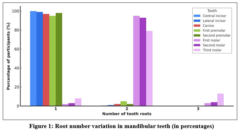 | Figure 1: Root number variation in mandibular teeth (in percentages) |
Figures 2 and 3 show CBCT scans of the same patient, demonstrating bilateral two-rooted morphology of the lower premolars. On both the right and left sides of the mandible, the premolars clearly exhibit two distinct roots, indicating a symmetrical anatomical variation.
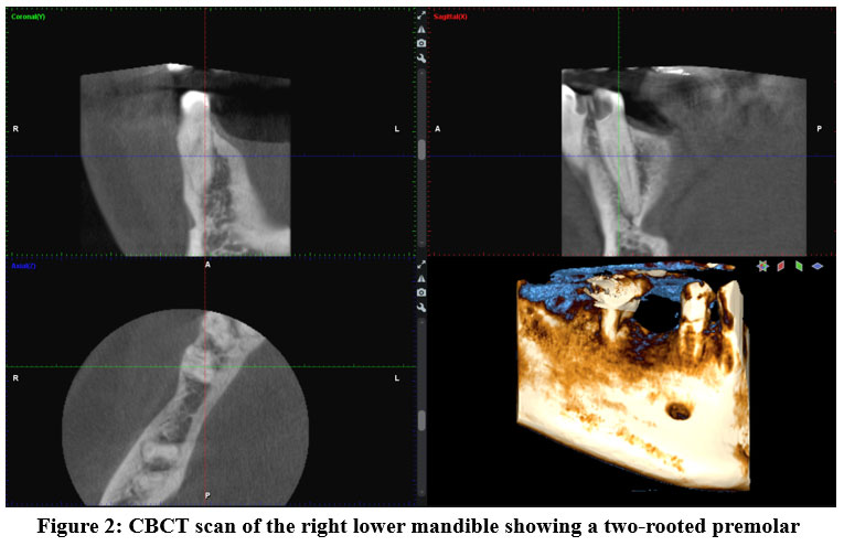 | Figure 2: CBCT scan of the right lower mandible showing a two-rooted premolar |
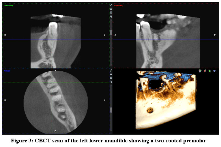 | Figure 3: CBCT scan of the left lower mandible showing a two-rooted premolar |
Discussion
All analysed lower central incisors were single-rooted, consistent with Krishnan et al.5 (Indian population), as well as several other studies.4,16,17 Lateral incisors were predominantly single-rooted, with two roots found in fewer than 1% of cases, supporting findings by Mashyakhy and Gambarini.17
Mandibular canines, in most cases, have one long and voluminous root, and since the small number of teeth analysed the percentage of 3.1% of lower canines with two roots is significant.16 Literature1 reports two-rooted lower canines in up to 10% of cases, including studies by Shrestha et al.4 and Magat and Uzun18. Pan et al.19 found 1.2% two-rooted canines in a Malaysian population, in line with our data.
Lower first premolars were mainly single-rooted, with 5% showing two roots, similar to previous studies.5–8,13,20,21 In an older study by Trope et al.22 two populations were analysed and a higher frequency of double-rooted premolars was observed in one population. In a study by Gündüz et al.,23 conducted on the Turkish population, out of a total of 988 analysed lower first premolars, 89.9% had one root, 9.7% had two roots, and 0.4% had even three roots.
Mandibular second premolars were also mostly single-rooted, with rare two-rooted cases, consistent with literature.5–8,13,20 Some studies16,17,19 have found that all lower second premolars analysed are teeth that have only one root. As with the lower first premolars, the study by Almehrzi et al.7 reported that the lower second premolars are single-rooted teeth with a lower proportion of fused shapes (the letter “C” shape). Gündüz et al.,23 out of a total of 974 teeth studied, also found lower second premolars with three roots in 0.3% of cases.
The lower first molars are mostly two-rooted teeth, but they can also have a supernumerary root (radix entomolaris or radix paramolaris), as shown by the results of our study. Similar results have been reported by other studies.14–16,24 Analyzing 1174 lower first molars, Qiao et al.9 reported in their study that 23.85% of the teeth had a supernumerary roots.
When it comes to lower second molars, this study found that these teeth are most often two-rooted, but in 10% of cases they can have one or three roots, which is largely confirmed by previous study.11,16,25,26 The study by Najžar-Fleger and Šutalo14 also recorded a fourth root. Kato et al.27 gave different results by analysing 22 lower second molars, in which the highest percentage had one root (64%), a slightly lower percentage had two roots (32%), and the lowest percentage had three roots (0.35%).
The lower third molars are the most variable teeth missing in the subjects in this study. The majority are two-rooted third molars, while the remaining proportion is single-rooted and three-rooted teeth, which is consistent with the available data from previous study.12
Taking into account the limiting factors of this study, namely the small number of samples and data processing from only two departments of the Clinical Hospital of Split, recorded over a period of one year, there is a need for further analysis of as many samples (CBCT scans) as possible throughout the Republic of Croatia.
Conclusion
Dental practitioners must be familiar with possible morphological variations in tooth roots to ensure successful endodontic therapy. Based on the results of the study, reading 114 CBCT images, it is concluded that the number of roots of all teeth in the lower jaw is largely consistent with the data from the literature on the stated number of roots. All three hypotheses were confirmed:
Lower incisors, canines and premolars are single-rooted teeth in the highest percentage.
Molars are mostly double-rooted teeth.
The most common type of variation is the supernumerary root of the lower third molar.
Acknowledgement
The authors thank the Split Clinical Hospital Center for the CBCT data provided.
Funding Sources
The author(s) received no financial support for the research, authorship, and/or publication of this article.
Conflict of Interest
The authors do not have any conflict of interest.
Data Availability Statement
This statement does not apply to this article.
Ethics Statement
This retrospective study was approved by the Institutional Ethics Committee of the Split Clinical Hospital (Class: 500-03/21-01/34; Reg. No.: 2181-147-01/06/M.S.-20-02). The requirement for informed consent was waived due to the retrospective design and use of fully anonymized CBCT data.
Informed Consent Statement
This study did not involve human participants, and therefore, informed consent was not required
Clinical Trial Registration
This research does not involve any clinical trials.
Permission to Reproduce Material from other Sources
Not Applicable
Author Contributions
Lucija Kustra: Conceptualization, Methodology, Data Collection, Analysis, Writing – Original Draft.
Ivana Medvedec Mikic - Analysis, Writing – Review & Editing.
Kristina Gorseta - Conceptualization, Writing – Review & Editing, Supervision.
References
- Karobari MI, Alam BF, Bashir R, Fahim MF, Mirza MB, Noorani TY. Bibliometric analysis: Root and root canal morphology using cone-beam computed tomography. Clin Exp Dent Res. 2023;9(6):1156-1168. doi:10.1002/cre2.801
CrossRef - Patel S, Brown J, Pimentel T, Kelly RD, Abella F, Durack C. Cone beam computed tomography in Endodontics - a review of the literature. Int Endod J. 2019;52(8):1138-1152. doi:10.1111/iej.13115
CrossRef - Krishnan G, E SM, Rangappa A, Rangaswamy V, Murthy CS, Kumar N N. Radicular Dentin Thickness and Root Canal Morphology of Mandibular Incisors in Indian Subpopulation Using Cone Beam Computed Tomography. Cureus. 2024;16(11):e73355. doi:10.7759/cureus.73355
CrossRef - Shrestha K, Shubham S, Ahmed S, Gautam V. Variations in the Root Form and Root Canal Morphology of Permanent Mandibular canine. J Nepal Health Res Counc. 2024;21(3):463-466. doi:10.33314/jnhrc.v21i3.4707
CrossRef - Alsofi L, Al-Habib M, Zahran S, et al. Three-dimensional evaluation of root canal morphology in mandibular premolars of Saudi individuals: a CBCT study. Libyan J Med. 2025;20(1).
CrossRef - Al-Zubaidi SM, Almansour MI, Alshammari AS, et al. Root and Canal Morphology of Mandibular Premolars in a Saudi Subpopulation: A Cone-Beam Computed Tomography Study. Int J Dent. 2022;2022:7. doi:10.1155/2022/4038909
CrossRef - Almehrzi H, Khawajah S, Alharbi N, Abed RE, Jamal M. Evaluation of the Root and Canal Morphology of Maxillary and Mandibular Premolars in an Emirati Sub-Population. Int Dent J. Published online 2025:1-10. doi:10.1016/j.identj.2025.02.001
CrossRef - Martins JNR, Tummala S, Nallapati S, et al. Population-Specific Anatomical Variations in Premolar Root Canal Systems: A Cross-Sectional Cone-Beam Computed Tomography Study of Jamaican and Portuguese Subpopulations. Dent J. 2025;13(2):2. doi:10.3390/dj13020050
CrossRef - Qiao X, Zhu H, Yan Y, et al. Prevalence of middle mesial canal and radix entomolaris of mandibular first permanent molars in a western Chinese population: an in vivo cone-beam computed tomographic study. BMC Oral Health. 2020;20(1):224. doi:10.1186/s12903-020-01218-z
CrossRef - Yang L, Han J, Wang Q, Wang Z, Yu X, Du Y. Variations of root and canal morphology of mandibular second molars in Chinese individuals: a cone-beam computed tomography study. BMC Oral Health. 2022;22(1):274. doi:10.1186/s12903-022-02299-8
CrossRef - Guo Q, Wang Q, Yang Y, Guo D. Root and root canal morphology of mandibular second permanent molars in the Gansu province population: A CBCT study. Aust Endod J. 2023;49(S1):27-32. doi:10.1111/aej.12692
CrossRef - Džankovi? A, Mahmutovi? A, Kora? S, et al. Istraživanje morfologije kanala korijena tre?ih kutnjaka u populaciji Bosne i Hercegovine. Acta Stomatol Croat. 2024;58(3):255-266. doi:10.15644/asc58/3/6
CrossRef - Šutalo J, Njemirovskij V. Morfološke karakteristike korjenova gornjih i donjih pretkutnjaka. Acta Stomatol Croat. 1980;14(1-2):23-28.
- Najžar-Fleger D, Šutalo J. Morfološke varijabilnosti korjenova prvog i drugog donjeg kutnjaka. Acta Stomatol Croat. 1984;18(3):203-209.
- Šutalo J, Najžar-Fleger D. Morfološke varijabilnosti korjenova prvog i drugog donjeg kutnjaka: prekobrojni korjenovi. Acta Stomatol Croat. 1984;18(2):119-125.
- Maluf TC, Bueno CE, Pelegrine RA, et al. Analysis of morphology and symmetry of the root canal system of incisors, premolars and mandibular molars using CBCT. Acta Odontológica Latinoam. 2024;37(1):25-33. doi:10.54589/aol.37/1/25
CrossRef - Mashyakhy M, Gambarini G. Root and Root Canal Morphology Differences Between Genders: A Comprehensive in-vivo CBCT Study in a Saudi Population. Acta Stomatol Croat. 2019;53(3):213-246. doi:10.15644/asc53/3/5
CrossRef - Magat G, Uzun S. Evaluation of root and root canal morphology of mandibular and maxillary canine teeth in Turkish subpopulation by cone beam computed tomography with using two classification systems. BMC Oral Health. 2024;24(1):1499. doi:10.1186/s12903-024-05252-z
CrossRef - Pan JYY, Parolia A, Chuah SR, Bhatia S, Mutalik S, Pau A. Root canal morphology of permanent teeth in a Malaysian subpopulation using cone-beam computed tomography. BMC Oral Health. 2019;19(1):14. doi:10.1186/s12903-019-0710-z
CrossRef - Nikoli? M, Miti? A, Gaši? J, et al. Varijacije broja korenova, kanala i dužine zuba kod prvih premolara. Dent J Serbia. 2014;49:37-41. doi:10.5937/gads1449037N
CrossRef - Pedemonte E, Cabrera C, Torres A, et al. Root and canal morphology of mandibular premolars using cone-beam computed tomography in a Chilean and Belgian subpopulation: a cross-sectional study. Oral Radiol. 2018;34(2):143-150. doi:10.1007/s11282-017-0297-5
CrossRef - Trope M, Elfenbein I, Tronstad L. Mandibular premolars with more than one root canal in different race groups. J Endod. 1986;12(8):343-345.
CrossRef - Gündüz H, Özlek E. Evaluation of root morphology and root canal configuration of mandibular and maxillary premolar teeth in Turkish subpopulation by using cone beam computed tomography. East J Med. 2022;27(3):465-471. doi:10.5505/ejm.2022.66743
CrossRef - Alazemi HS, Al-Nazhan SA, Aldosimani MA. Root and root canal morphology of permanent mandibular first and second molars in a Kuwaiti population: A retrospective cone-beam computed tomography study. Saudi Dent J. 2023;35(4):345-353. doi:10.1016/j.sdentj.2023.03.008
CrossRef - Al-Qudah AA, Awawdeh LA. Root and canal morphology of mandibular first and second molar teeth in a Jordanian population. Int Endod J. 2009;42(9):775-784. doi:10.1111/j.1365-2591.2009.01578.x
CrossRef - Gomez F, Brea G, Gomez-Sosa JF. Root canal morphology and variations in mandibular second molars: an in vivo cone-beam computed tomography analysis. BMC Oral Health. 2021;21(1):424. doi:10.1186/s12903-021-01787-7
CrossRef - Kato A, Hishikawa T, Inagaki K, Yamamoto G, Mitani A, Honda M. Evaluation of root morphology of maxillary and mandibular second molars lost due to periodontitis. J Periodontal Res. 2020;55(5):753-761. doi:10.1111/jre.12764
CrossRef


