.jpg)

A Case Report of Oral Pigmentation - Post-Inflammatory or Psoriasiform Lesions
Sujatha G.P1*  , Kalavathi G.P 2 and Ashok l1
1Department of Oral medicine and radiology , Bapuji dental college and hospital, Davangere, India .
2Department of General Medicine, Karawar instituite of medical sciences, Karawar, Uttarkannada India .
http://dx.doi.org/10.12944/EDJ.02.01.08
Intraoral pigmentation occurs in various physiologic and pathologic conditions. Many of these are the manifestations of systemic diseases that too dermatologic conditions in specific. Here with reporting a case of intraoral brownish pigmentation in a young adult female who was treated for mild dermal lesions of psoriasis which was reported to be either post inflammatory pigmentation or psoriasiform oral manifestations. The pattern of mucosal pigmentation is nonspecific and there is lack of recent research regarding etiopathogenesis.
Oral Pigmentation; Post-inflammatory Pigmentation; Psoriasiform lesions
Copy the following to cite this article:
Sujatha G. P, Kalavathi G. P, Ashok l. A Case Report of Oral Pigmentation - Post-Inflammatory or Psoriasiform Lesions. Enviro Dental Journal 2022; 2(1). DOI:http://dx.doi.org/10.12944/EDJ.02.01.08
Copy the following to cite this URL:
Sujatha G. P, Kalavathi G. P, Ashok l. A Case Report of Oral Pigmentation - Post-Inflammatory or Psoriasiform Lesions. Enviro Dental Journal 2022; 2(1). Available From: https://bit.ly/3IMXU1F
Download article (pdf) Citation Manager

Introduction
Intraoral pigmentation is one of the most common clinical conditions, despite pose a challenging situation to diagnose often. Post inflammatory pigmentation is one such entity where in elucidation of etiopathogenesis remain unanswered. Herewith reporting a common case of intraoral brownish pigmentation that ignites a new interrogation.
Case-report
A 24 year old female patient presented with asymptomatic brownish discolouration of oral mucosa since 1 month. The discoloration was gradual in on set, was initially smaller in size and gradually increased to the present size. Patient was diagnosed with milder variant of Psoriasis over both the elbows 2 months ago by a dermatologist and treated with topical steroid cream( triamcinolone acetonide-0.1%) application and short term systemic steroid therapy (90mg/day for first 3days tapered to 30mg /day for 9 days) and also gives history of appearance of oral lesion after the resolution of the skin lesions. Dental and family history were non-contributory.
Extraoral manifestation revealed Small scaly popular eruptions over the skin of right elbow. [Figure No 1]
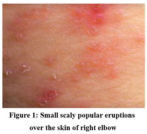 |
Figure 1: Small scaly popular eruptions over the skin of right elbow. |
Intraoral examination revealed the multiple diffuse brownish to black pigmentation of oral mucosa chiefly over hard palate, attached gingiva, tongue & buccal mucosa.(FIG No. 2-5) The lesions are irregular in shape with well-defined margins. On palpation the lesion was non tender, non-scrapable, do not disappear on stretching.
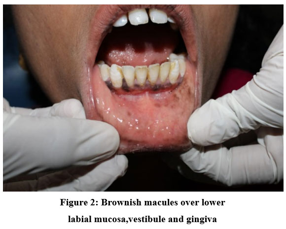 |
Figure 2: Brownish macules over lower labial mucosa,vestibule and gingiva |
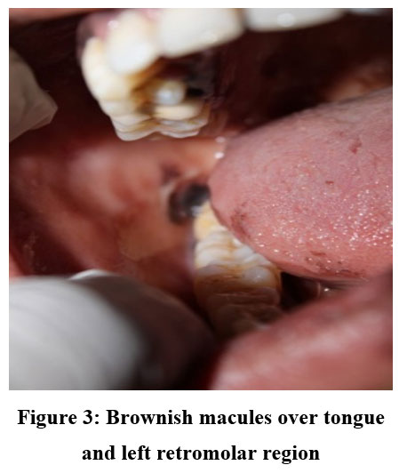 |
Figure 3: Brownish macules over tongue and left retromolar region. |
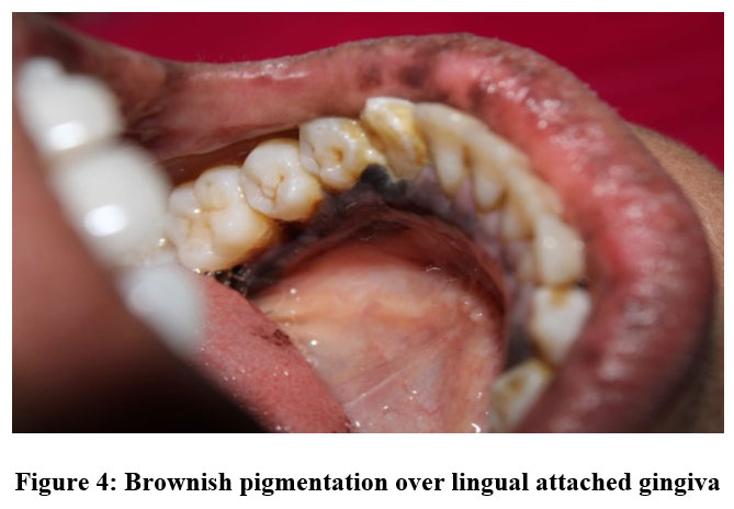 |
Figure 4: Brownish pigmentation over lingual attached gingiva. |
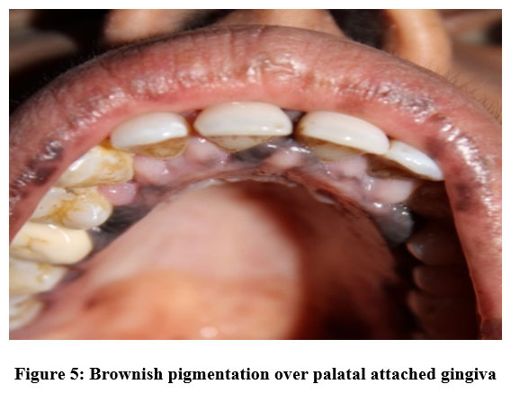 |
Figure 5: Brownish pigmentation over palatal attached gingiva. |
Provisional diagnosis of endogenous Intraoral pigmentation and differential diagnosis of post inflammatory pigmentation, unnoticed oral manifestation of psoriasis or drug induced was given.
As patient was uncooperative for further investigations, as the appearance of the mucosal lesion was observed secondary to skin lesion and corresponded with available reports the lesion could be secondary to psoriasis.
Discussion
Colour of the normal oral mucosa is pale pink to reddish depending on the vascularity, thickness of the epithelium, amount of melanin pigment deposition from melanocytes and degree of keratinisation. Pigmentations of oral mucosa are result of increased production of melanin and augmented number of melanocytes and deposition of exogenous materials.1.
Melanin, an endogenous non hematogenous pigment secreted by melanocytes in basal layer of oral epithelium & is transferred to adjacent cells via melanosomes. Neural crest derivatives or nevus cells also synthesize and cause melanin pigmentation. Blue,brown and black discoluration of tissues are influenced by the region and amount of melanin pigmentation.2,3, 4
Healthy individuals like dark skinned African population exhibit aboutmelanin pigmentation 100%compareted to 30% - 98% of Asians.2 Oral pigmentation can be classified as physiologic and pathologic. Pathologic pigmentation can be either exogenous or endogenous based upon etiologic factors. Exogenous pigmentation occurs due to drugs, smoking/smokeless use of tobacco habbit, amalgam tattoo or heavy metals induced. Endogenous pigmentation commonly seen in endocrine disorders, systemic disease or syndromes, chronic irritation or microtrauma, microbial infections, reactive or neoplastic disorders.2,3, 4
Post-inflammatory pigmentation is less well studied type of pigmentation which occurs as a consequence of chronic inflammatory or refractory oral mucosal diseases, particularly oral lichen planus. Dark-skinned personelles frequently exhibit diffuse brownish to black pigmentary areas adjacent to reticular or erosive lesions of lichen planus.It is also quoted that once the healing of the lesion manifest as pigmentation. Halder et al. reported that pathogenesis of post-inflammatory pigmentation remains imprecise. Histological sections reveal increased amount of melanin laid by melanocytes and superficial connective tissue showing accumulation of melanin laden macrophages. 3, 6,7
Various drugs like hormones, oral contraceptives, chemotherapeutic such as cyclophosphamide, busulfan, bleomycin & fluorouracil, transquilizers, antimalarials like clofazamine, chloroquine, amodiaquine, commonly used anti-microbials like minocycline, antivirals such as zidovudine and antifungals like ketaconazole very commonly known to iduce pigmentation in the tissues and oral mucosa. 9
Palatal mucosa and gingiva are most commonly affected sites. Along with mucosal discoloration,teeth in adults & children may be bluish gray due to minocycline/tetracycline therapy. Drug-related pigmentation occurs as of drug itself or metabolites of drug deposition in dermis & epidermis, increased melanin accumulation with or without upsurge in number of melanocytes. Drug-induced post-inflammatory pigmentaion of oral mucosa is obsreved specifically if drugs persuade an oral lichenoid reaction & bacterial metabolism, alone or in combination.2-10
Psoriasis is primarily a papulosquamous dermatologic disease that manifest with skin lesions of variable morphology, distribution, severity, and course. Patients with Psoriasis complain of intense pruritus or burning.Skin lesions are characterised by papules and plaques with scaling characteristically disseminated symmetrically over scalp, elbows, knees, lumbosacral area, & in body folds. Koebner’s phenomenon ie occurance of lesions at site of trauma or injury, is seen in Psoriasis. A generalised exfoliative erythroderma.is thefeature of psoriasis which is progressive or uncontrolled. This can also result in Nail involvement and psoriatic arthritis (PsA). Infrequently psoriasis may comprise oral mucosa or tongue. Features of tongue include discrete circumscribed white and red patches with circinate white yellow border and changing pattern that assume distinct annular patterns in map like fashion, hence the name geographic tongue. Till now there are no reports of associated oral pigmentation. 8.9 &10 The present case report could be the oral counter part of dermal psoriatic lesions.
Conclusion
Black and Brown oral mucosal pigmentation is seen in various physiologic conditions to life-threatening diseases. Need of an hour is early identification of unusual pigmentation of oral mucosa by Oral physicians through appropriate clinical history regarding the onset, duration, & existence of local or systemic symptoms. Some lesions can be diagnosed clinically or by laboratory testing and biopsy is indicated in suspicious lesions.
Conflicts of interest
There is no conflicts of interest.
Funding Sources
There is no funding Source.
References
- C Sreeja , K. Ramakrishnan, D. Vijayalakshmi, M. Devi, I. Aesha, and B. Vijayabanu Oral pigmentation: A review. J Pharm Bioallied Sci. 2015 Aug; 7(Suppl 2): S403–S408
CrossRef - 2 Anil Kumar N, Divya P. Adverse drug effects in mouth. International Journal Of Medical And Applied Sciences. 2015;4:82–913
- Greenberg M, Glick M. Burkets oral medicine diagnosis and treatment. 10th ed. Hamilton, Ontario: B. C. Decker; 2003. pp. 126–36
- Molly S. Rosebush, Ashleigh NBriody, Kitrina G. Cordell:Black and Brown: Non-neoplastic Pigmentation of the Oral Mucosa. Head and Neck Pathology (2019) 13:47–55
CrossRef - R M Halder,P K Nootheti.Ethnic skin disorders overview J Am Acad Dermatol,48 (2003)
CrossRef - Ayeisha Umaira Durga Prasad Mohammed Alsakran Altamimi A review:Diagnosis of oral pigmentations and malignant transformations. Singapore Dental Journal Volume 35, December 2014, Pages 39-46.
CrossRef - Hatch CL. Pigmented lesions of the oral cavity. Dent Clin North Am. 2005;49:185–201. ix. [PubMed] [Google Scholar
CrossRef - Report:Psoriasis: epidemiology, clinical features, and quality of life R G B Langley, G G Krueger, C E M Griffiths Ann Rheum Dis 2005;64(Suppl II):ii18–ii23. doi: 10.1136/ard.2004.033217
CrossRef - Pathophysiology, Clinical Presentation, and Treatment of Psoriasis:A Review.April W. Armstrong, MD, MPH1; Charlotte Read, MBBS, BSc1,2 JAMA. 2020;323(19):1945-1960.
CrossRef - Neville BW, Damm DD, Allen CM, Bouquot JE. Oral and Maxillofacial Pathology. 3rd ed. St. Louis: Saunders Elsevier Publications: Elsevier Publications; 2009. pp. 308–13.